Download presentation
Presentation is loading. Please wait.

1
AH Biology: Unit 1 Communication Within Multicellular Organisms

2
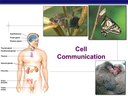
Communication within multicellular organisms
General principles. Hydrophobic signals and control of transcription. Hydrophilic signals and transduction.

3
In animals communication is mediated by nervous transmission and hormonal secretion.
Compare and contrast the two modes of communication. Challenge students to name the glands shown in the diagram: 1. pineal; 2. pituitary; 3. thyroid; 4. thymus; 5. adrenal; 6. pancreas; 7. ovary; 8. testis

4
Nervous communication Hormonal communication
Nature of signal Electrical impulses and extracellular signalling molecules Extracellular signalling molecules Transmission of signal Along the axons of neurons Through the bloodstream Target cells Any cells with connections to neurons (effectors) Almost any cells in the body Time for response to occur Faster Slower Duration of response Transient Longer lasting Extent of response Localised Widespread As an analogy: a person on a desert island can send messages to the outside world in bottles (hormonal communication) or use a telephone (nervous communication). For extent of response: one hormone can act on many spatially separate organs: a nerve impulse results in the contraction of one muscle in one part of the body or the secretion of a chemical from one gland in the body.
 Almost any cells in the body. Time for response to occur. Faster. Slower. Duration of response. Transient. Longer lasting. Extent of response. Localised. Widespread. As an analogy: a person on a desert island can send messages to the outside world in bottles (hormonal communication) or use a telephone (nervous communication). For extent of response: one hormone can act on many spatially separate organs: a nerve impulse results in the contraction of one muscle in one part of the body or the secretion of a chemical from one gland in the body.")
5
Coordination is important for homeostasis
Brainstorm all the physiological variables that must be maintained within a narrow range of values.

6
Coordination allows integrated homeostatic responses to be made.
Disturbances Coordinated responses Error- correcting mechanisms Controlled system Monitoring centres Error signal Set point values

7
Coordination of responses allows animals to cope with physiological stress, eg a human doing exercise. . . Discuss what challenges exercise presents and the coordinated homeostatic responses that would be required.

8
Exercise Cardiovascular challenge Ventilatory challenge
Metabolic challenge Thermoregulatory challenge Osmoregulatory challenge What responses would be needed to meet these challenges? No need to mention specific hormones just yet. Cardiovascular challenge: increased blood flow to muscles, decreased blood flow to gut, increased heart rate and force of contraction (to raise arterial blood pressure to compensate for decrease in total peripheral), constriction of veins to increase rate of venous return so heart does not run out of blood to pump. Respiratory challenge: Increased rate of ventilation (actually an anticipatory response), dilation of bronchi. Metabolic challenge: More glucose must be supplied to respiring tissue, increased glycogenolysis in muscles and liver, and increased gluconeogenesis in liver, release of free fatty acids from adipose tissue. All responses are promoted by adrenaline. Thermoregulatory challenge: Heat loss must increase. Increased sweating and vasodilation of skin blood vessels. Osmoregulatory challenge: Reduced volume of urine produced, increased reabsorption of salt.
, constriction of veins to increase rate of venous return so heart does not run out of blood to pump. Respiratory challenge: Increased rate of ventilation (actually an anticipatory response), dilation of bronchi. Metabolic challenge: More glucose must be supplied to respiring tissue, increased glycogenolysis in muscles and liver, and increased gluconeogenesis in liver, release of free fatty acids from adipose tissue. All responses are promoted by adrenaline. Thermoregulatory challenge: Heat loss must increase. Increased sweating and vasodilation of skin blood vessels. Osmoregulatory challenge: Reduced volume of urine produced, increased reabsorption of salt.")
9
Extracellular signalling
Signalling cells Specific signalling molecules released as a result of a change in internal state Signalling molecules carried to target cells Target cells Arrival of signalling molecules at target cells is linked to a change in the internal state of the cells (cell response)
")
10
Extracellular signalling
Signalling cells Specific signalling molecules released as a result of a change in internal state Signalling molecules carried to target cells Target cells (may also act as signalling cells) Arrival of signalling molecules at target cells is linked to a change in the internal state of the cells (cell response)
 Arrival of signalling molecules at target cells is linked to a change in the internal state of the cells (cell response)")
11
Different cell types produce specific signalling molecules.
Challenge students to name the hormones produced by the glands shown and describe what their target cells are and what is produced by the nerves (neurotransmitters). Pineal gland: melatonin (regulates circadian rhythms). Pituitary gland: ADH and GH. Thyroid gland: thyroxine. Thymus gland: thymosins (promote development of lymphocytes). Adrenal glands: adrenaline and aldosterone (salt retention). Pancreas: insulin and glucagon. Ovaries: estrogen and progesterone. Testes: testosterone.
. Pineal gland: melatonin (regulates circadian rhythms). Pituitary gland: ADH and GH. Thyroid gland: thyroxine. Thymus gland: thymosins (promote development of lymphocytes). Adrenal glands: adrenaline and aldosterone (salt retention). Pancreas: insulin and glucagon. Ovaries: estrogen and progesterone. Testes: testosterone.")
12
Spatial organisation of signalling molecules
Eukaryotic cell: 50 μm Distance: 1 nm μm mm m km Animal pheromones Pheromones are chemical signals that are used to transmit information between organisms. Note that the scale is non-linear. Hormones Neurotransmitters

13
How does a target cell ‘know’ that it should respond to a specific signal?

14
Cells can only detect and respond to signals if they possess a specific receptor.
Insulin Adrenaline Insulin receptor protein Adrenaline receptor protein

15
Different cell types may show a specific tissue response to the same signal.
Beta-receptor Adrenaline Beta-receptor Adrenaline Amylase release stimulated Glycogen breakdown stimulated Cell in mammalian salivary gland Cell in mammalian liver

16
Hydrophobic signals and control of transcription
One way of classifying extracellular signalling molecules is by hydrophobicity.

17
Action of hydrophobic signalling molecules
Hormone Altered rate of protein synthesis (long-lasting effects) Intracellular receptor protein Altered rate of gene transcription
 Intracellular receptor protein. Altered rate of gene transcription.")
18
Animation of regulation of transcription.
Hydrophobic signalling molecules can bind to nuclear receptors to regulate gene transcription. Animation of regulation of transcription.

19
Steroid hormones are hydrophobic signalling molecules.
Animation of mechanism of steroid hormone action.

20
The steroid hormone receptor proteins are transcription factors.
Inhibitory protein complex Inactive transcription factor Hormone-binding site Steroid hormone Active transcription factor DNA-binding site exposed

21
Thyroxine is a hydrophobic hormone that regulates the metabolic rate.
Why is thyroxine not classified as a carbohydrate, lipid or protein?

22
Thyroxine is released from the thyroid gland.

23
Thyroxine absent Thyroid receptor protein bound to DNA
Transcription of Na+/K+ ATPase gene inhibited

24
Action of thyroxine Thyroxine Receptor protein
undergoes conformational change Synthesis of Na+/K+ ATPase Transcription of Na+/K+ATPase gene

25
More Na+/K+ATPases in cell membrane
ATP degraded faster Increased metabolic rate Insertion into membrane Synthesis of Na+/K+ ATPase Transcription of Na+/K+ATPase gene

26
Hydrophilic signals and transduction

27
Hydrophilic ligands Molecules that bind to sites on target proteins (receptors) at the surface of cells to trigger signal transduction. Ligand binding triggers the receptor protein to undergo a conformational change.

28
Hydrophilic signal Reception + transduction Amplification
Second messenger Internal regulator Tissue-specific effectors Cell responses

29
Action of hydrophilic signalling molecules
Hormone (ligand) Receptor protein Similarity between mode of action = hormones bind to specific proteins. Hormone – receptor shapes are complementary. Hormones can only act on a target tissue if it possesses its specific receptors. Signal transduction Cell responses (short-lasting effects)
 Receptor protein. Similarity between mode of action = hormones bind to specific proteins. Hormone – receptor shapes are complementary. Hormones can only act on a target tissue if it possesses its specific receptors. Signal transduction. Cell responses. (short-lasting effects)")
30
Peptide hormones are short chains of amino acids.
ADH Insulin

31
Animation of action of acetylcholine.
Neurotransmitters are chemical signals released from nerve endings that alter the activity of target cells. Axon Neurotransmitter substance Synapse Location of receptors Animation of action of acetylcholine.

32
Hydrophilic signal transduction 2: receptors with kinase activity
Part of receptor that binds insulin (alpha-subunit) Part of receptor with kinase activity (beta-subunit) Insulin as example.
 Part of receptor with kinase activity (beta-subunit) Insulin as example.")
33
Hydrophilic signal transduction 1: G-protein cascade
Adenylate cyclase enzyme - + Stimulatory G-protein Inhibitory G-protein + Membrane channels + pumps, microtubules, histones, specific enzymes cAMP (second messenger) + Protein kinase A Animation of G-protein activation.
 + Protein kinase A. Animation of G-protein activation.")
34
P P P P P 1. Insulin binds to receptor
2. Kinase enzyme phosphorylates itself (autophosphorylation) P P P 4. Phosphorylated IRS-1 acts on effectors to trigger cell responses Insulin as example. 3. Receptor phosphorylates insulin receptor substrate (IRS-1) P Animation of protein kinase activity triggered by adrenaline and tyrosine kinase activity.
 P. P. P. 4. Phosphorylated IRS-1 acts on effectors to trigger cell responses. Insulin as example. 3. Receptor phosphorylates insulin receptor substrate (IRS-1) P. Animation of protein kinase activity triggered by adrenaline and tyrosine kinase activity.")
35
Insulin regulates the glucose concentration of the blood
Beta-cells in pancreas release more insulin Insulin transported in blood ADH acts on adipose, liver and muscle cells Change detected More glucose is taken up by cells Blood glucose concentration falls Blood glucose concentration rises Blood glucose concentration at set point

36
Action of insulin on fat and muscle cells
GLUT4 Note that GLUT4 is an insulin-dependent glucose transporter found in adipose and striated muscle tissue. Liver has GLUT2 insulin-independent glucose transporter. Liver is still sensitive to insulin. Insulin stimulates liver to convert glucose into glycogen. Animation of insulin action.

37
GLUT4 recruitment is also induced by exercise.

38
Diabetes mellitus A disease caused by defects in the insulin signalling system. Two types of diabetes mellitus are recognised. What are the general symptoms of diabetes mellitus? Etymology: Diabetes means excessive urine/flow run through/siphon. Mellitus means sweet. Diabetes has triad of symptoms: polyuria, polydypsia and polyphagia (excessive urination, thirst and hunger).
.")
39
Type 1: Insulin-dependent diabetes
Type 2: Non-insulin-dependent diabetes Cause Destruction of beta-cells in pancreas by immune system Exact cause unknown Obesity is a risk factor Usual age of onset Childhood Adulthood Nature of defect Pancreas does not produce any insulin Target cells develop insulin resistance Loss of receptor function Treatment Daily insulin injections and management of diet to control blood glucose concentration Eat less sugar and saturated fat Regular exercise Medication to lower blood glucose concentration Type 1 diabetes mellitus autoimmune reaction has a genetic component. Type 2 also has a genetic component.

40
Global prevalence of diabetes mellitus
The majority of cases are type 2 diabetes mellitus. Numbers are millions!

41
Review of diabetes mellitus
Animation of insulin production and type 1 diabetes mellitus. Basic animation of type 2 diabetes mellitus. Animation of type 2 diabetes mellitus.

42
Terrestrial vertebrates require mechanisms for conserving water
Thank goodness I can make ADH!

43
ADH regulates the body’s water balance
Pituitary gland releases more ADH ADH transported in blood ADH acts on kidney collecting ducts Change detected More water reabsorbed into blood Less urine made Blood water concentration falls Blood water concentration rises Blood water concentration at set point

45
Mechanism of action of ADH
Lumen of collecting duct Collecting duct cell Blood 2. ADH receptor H2O 1. ADH 5. Fusion of vesicles containing AQP2 water channel proteins Rate-limiting step in water reabsorption is uptake across the luminal membrane. The water permeability of the basolateral membrane is always high. 4. Protein phosphorylation 3. Activation of protein kinase A

46
Aquaporins are protein channels that allow efficient transmembrane movement of water.
Animation of water movement through an aquaporin channel.

47
Diabetes insipidus Disease in which the water conservation mechanism of the kidneys fails. What could the nature of the failure be? What would the symptoms of diabetes insipidus be?

48
The two types of diabetes insipidus
Central diabetes insipidus: insufficient ADH is produced. Nephrogenic diabetes insipidus: cells in the lining of the collecting duct are unable to respond to ADH. Compare these two types of diabetes insipidus to the two types of diabetes mellitus. Nephrogenic diabetes insipidus can be caused by mutations to the genes coding for the ADH receptor, signalling proteins or the AQP2 channel protein.

49
Possible causes of diabetes insipidus
Lumen of collecting duct Collecting duct cell Blood ADH receptor AQP2 ADH Note that non-functional protein kinase A would have wide-ranging effects. Phosphorylated target proteins Protein kinase A

50
Symptoms of diabetes insipidus
Excessive thirst. Production of large quantities of dilute urine (‘insipidus’ = lacks flavour). Compare these two types of diabetes insipidus to the two types of diabetes mellitus.
. Compare these two types of diabetes insipidus to the two types of diabetes mellitus.")
51
Overview of the action of ADH

Similar presentations


 2. hormones – islets of Langerhans 1 – 2% of pancreas are the islets.>")